In medical terms elbow is known as cubitus. It is a joint formed by humerus, radius and ulna bones It is a synovial joint of hinge type.
ARTICULAR SURFACE
There are two articular surfaces:
Upper Articular Surface – Formed by the capitulum and the trochlea of lower end of the humerus.
Lower Articular Surface – Formed by the upper surface of the head of the radius and trochlear notch of the ulna.

N.B – The trochlear notch of ulna is formed by the upper surface of the coronoid process and anterior surface of the olecranon process. The upper end of radius is circular in outline and slightly depressed in the center
LIGAMENTS
Capsular Ligament (Joint Capsule)
It is a fibrous sac enclosing the joint cavity. The inner surface of the capsule is lined by the synovial membrane. It is lax and thin anteriorly to allow the flexion and extension movements and is strengthened on either side by the collateral ligaments.
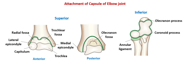
Attachment:
Above – It is attached to the medial epicondyle, margins of coronoid, radial and olecranon fossa, and lateral epicondyle.
Below – It is attached to the anterior and medial margin of coronoid process and upper and medial margin of olecranon process of ulna, and the annular ligament ( is not attached to radius).
Medial ligament (Ulnar collateral ligament)
- Triangular in shape.
- Extends from medial epicondyle of humerus to the upper end of ulna.
- Consists of three parts – anterior , inferior and posterior.

Attachment:
- Superiorly – It is attached is to the medial epicondyle.
- Inferiorly – The anterior thick band is attached to the medial margin of coronoid process. The posterior band is attached to the medial margin of olecranon process and the inferior thick transverse part extends from olecranon to coronoid process.
- It is related to the ulnar nerve.
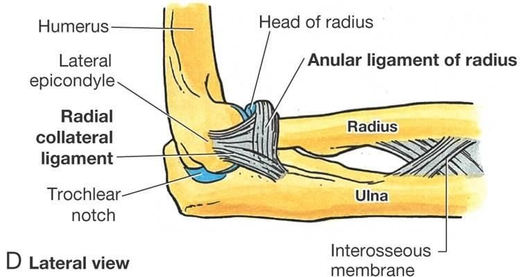
Lateral ligament (Radial collateral ligament)
It extends from lateral epicondyle of the humerus to the annular ligament with which it blends.
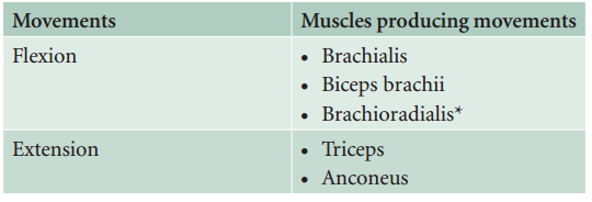
MOVEMENTS
BLOOD AND NERVE SUPPLY
Blood Supply:
Arterial anastomosis around the elbow formed by the branches of brachial, radial, and ulnar arteries.
Nerve supply:
By branches from musculocutaneous, medial, ulnar and radial nerves.
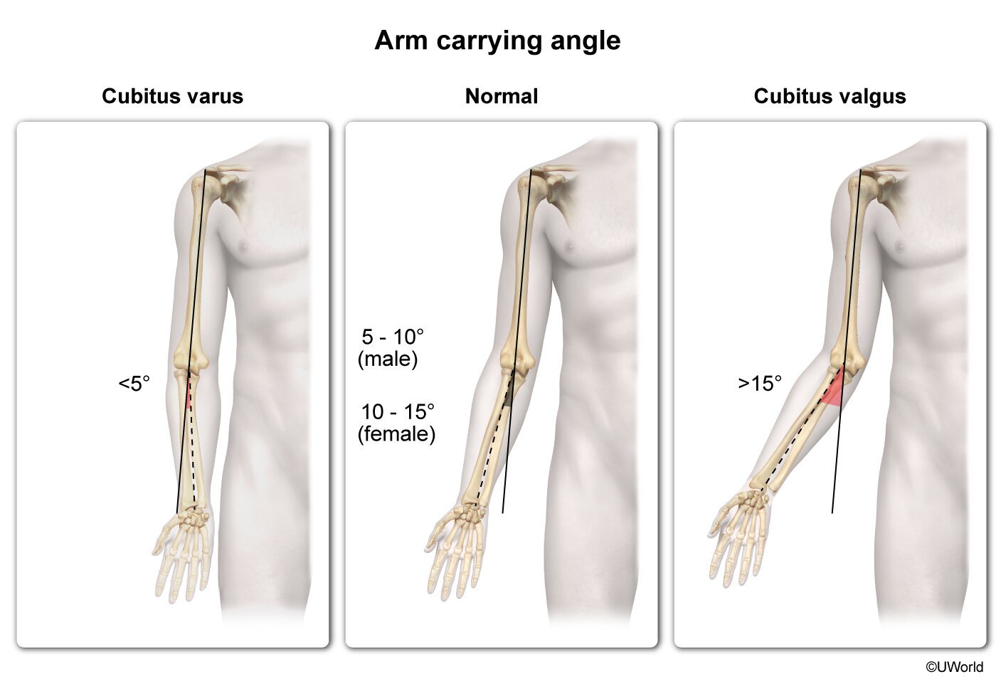
CARRYING ANGLE
When elbow is extended, the arm and forearm do not lie in a straight line, rather forearm is deviated laterally at 13° angle. So, it can be defined as angle between long axis of arm and forearm.
- Greater in shorter person when compared to taller person.
- This angle is greater in throwing athletes.
- Greater in females
Cubitus Varus – Less than 5%
Cubitus Valgus – More than 15%
CLINICAL ANATOMY
Elbow effusion
The distension of elbow joint due to effusion within its cavity occurs posteriorly because capsule of the joint is thin posteriorly and covering fascia is also thin. The joint is aspirated by inserting a needle on the posterolateral side, above the head of radius with elbow at the right angle.

Nursemaid’s elbow/pulled elbow
Occurs in preschool children when forearm is pulled suddenly in pronation. Head of radius come out of annular ligament and elbow is kept slightly flexed and pronated.

Tennis elbow (Lateral epicondylitis)
Pain and tenderness over lateral epicondyle of humerus with pain during abrupt pronation. It occurs due to strain or tear of common extensor origin and inflammation of lateral epicondyle.

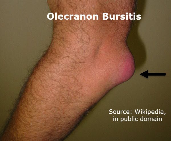
Students elbow
Round fluctuating panful swelling over olecranon. It occurs due to inflammation of subcutaneous olecranon bursa over the triangular posterior surface of olecranon process.
Golfer’s elbow (Medial epicondylitis)
Pain and tenderness over medial epicondyle of humerus. It occurs due to strain or tear of common flexor origin and inflammation of medial epicondyle.