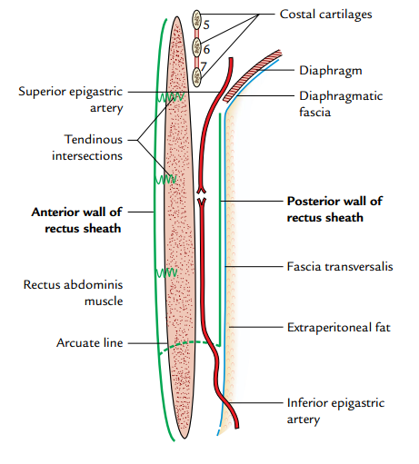
This the aponeurotic sheath covering the rectus abdominis muscle
Walls
Anterior –
- It’s a complete covering muscle from end to end
Posterior –
- Its incomplete. Deficient above the costal margin and below the arcuate line
Formation
The formation of rectus sheath differs from above downward as follows:
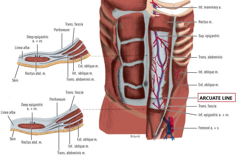
Above the level of costal margin
Anterior wall –
- It is formed by the aponeurosis of external oblique only.
Posterior wall –
- It is deficient and muscle lies directly on the 5th, 6th, and 7th costal cartilages
Between costal margin and arcuate line
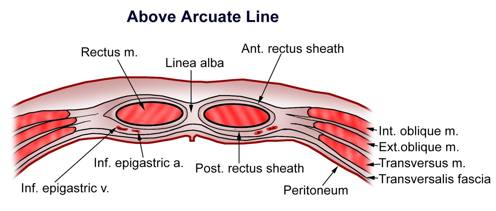
Anterior wall –
- It is formed by the fusion of aponeurosis of external oblique with the anterior lamina of aponeurosis of internal oblique.
Posterior wall –
- It is formed by the fusion of aponeurosis of transversus abdominis with the posterior lamina of aponeurosis of internal oblique.
Below the level of arcuate line
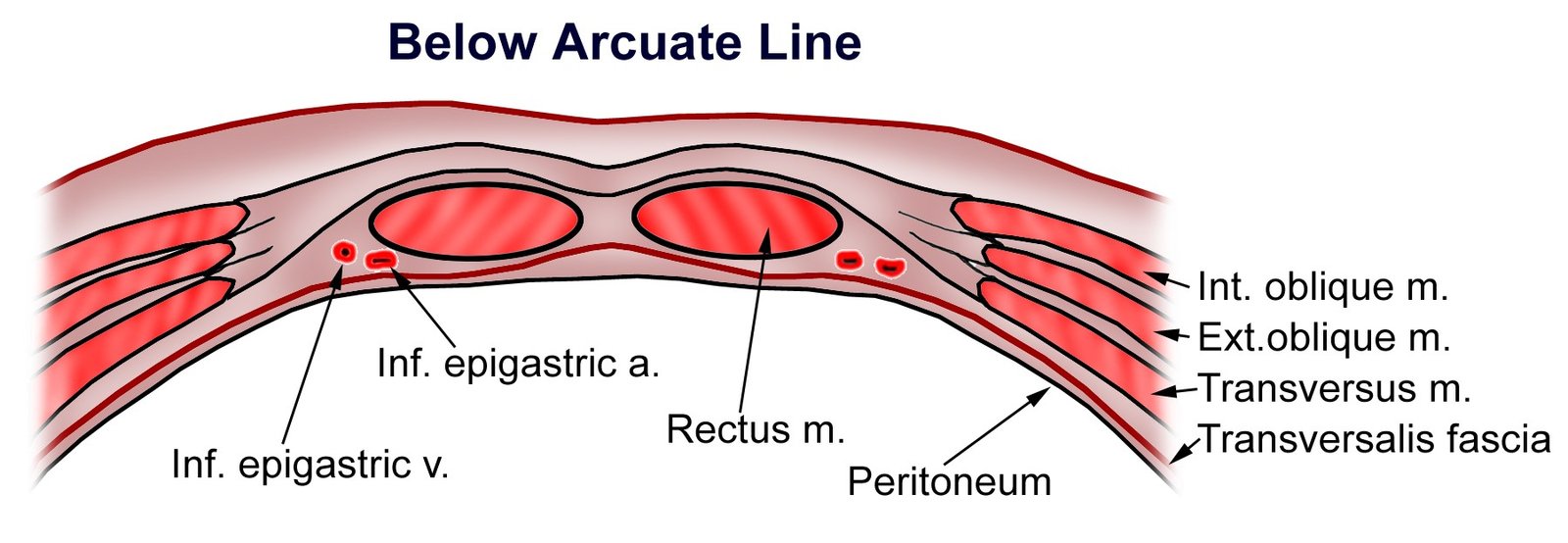
Anterior wall –
- It is formed by the aponeuroses of all the three flat muscles (the aponeuroses of transversus abdominis and internal oblique are fused but the aponeurosis of external oblique remains separate).
Posterior wall –
- It is deficient
- Directly rest on fascia transversalis
Arcuate line – Its location is half the distance from the pubic symphysis to the umbilicus. It is the demarcation where the internal oblique and transversus abdominis aponeuroses of the rectus sheath start to pass anteriorly to the rectus abdominis muscle, leaving only the transversalis fascia posteriorly. It is also where the inferior epigastric vessels perforate the rectus abdominis.
Contents of Rectus Sheath
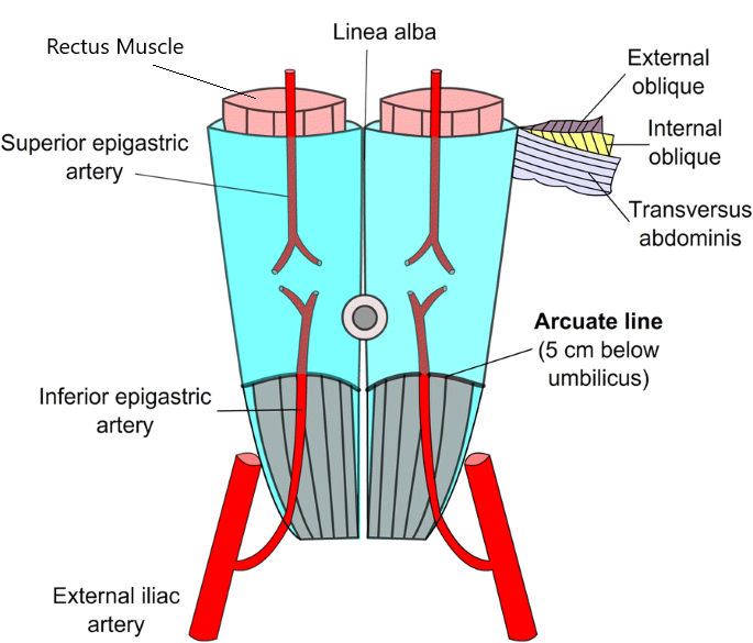
Muscles
- Rectus abdominis
- Pyramidalis (if present).
Arteries
- Superior epigastric artery branch of internal thoracic artery
- Inferior epigastric artery branch of external iliac artery.
Veins
Corresponding to the arteries.
Nerves
Terminal parts of lower six thoracic nerves
- Lower ‘5’ intercostal nerves (T7 – T11)
- Subcostal nerve.
Function of rectus sheath
- It checks the bowing of the rectus Abdominis muscle during contraction
- It maintains the strength of anterior abdominal wall
Applied Anatomy
- The pedicle flap of the upper part of the rectus abdominis muscle based on the superior epigastric artery is commonly used in the reconstructive surgery of the breast.
- Divarication of the recti (separation of the recti abdominis muscles): The separation of two rectus muscles usually occur in elderly multiparous woman with weak abdominal muscles. In this condition, the aponeuroses forming the rectus sheaths become excessively stretched, consequently when the patient coughs or strains, the recti separate widely and a hernial sac containing loops of intestine protrudes forward between the medial margins of the recti.
- Hematoma of rectus sheath: Sometimes the superior and inferior epigastric arteries are unduly stretched during a severe bout of coughing or in later months of pregnancy and ruptures if they are exposed to blunt trauma to the anterior abdominal wall leading to the formation of hematoma within the rectus sheath. Clinically, it presents as:
- Midline abdominal pain.
- Tender mass confined to one rectus sheath


[…] is made 1-1 ½ inch lateral and parallel to the midline, the anterior wall of rectus sheath is opened, the rectus abdominis is displaced laterally (to avoid injury to the nerves supplying […]