Plexus is a network of nerves or vessels in the body. The Brachial Plexus is a plexus of nerves formed by anterior/ ventral rami of lower four cervical (C5-8) and first thoracic (T1) spinal nerves. There may also a contribution from C4 (Prefixed Brachial Plexus) & T2 (Postfixed Brachial Plexus) can be seen.
Formation:
It consists of 4 components –
Roots
- Located in the neck deep to Scaleneus anterior muscle
- Constituted of anterior rami of C5-8 & T1 spinal nerves
Trunks
- Located in the neck between Scaleneus anterior & Scaleneus medius muscles
- Forms 3 trunks –
- Upper Trunk by joining C5 and C6 roots
- Middle Trunk by C7 root alone
- Lower Trunk by C8 and T1 roots
Divisions
- Located behind Clavicle
- Each trunk divides into 2 divisions forming total 6 divisions
Cords
- Located in the Axilla
- Forms 3 cords –
- Lateral Cord by Anterior divisions of Upper and Middle trunk
- Posterior Cord by Posterior divisions of all 3 (Upper, Middle, Lower) trunks
- Medial Cord by Anterior division of Lower Trunk alone

Branches:
From Roots
- Long Thoracic Nerve (C5,6,7) – Serratus anterior
- Dorsal Scapular Nerve (C5) – Rhomboids
From Trunks
- Suprascapular Nerve (C5,6) – Supraspinatus & Infraspinatus
- Nerve to Subclavius (C5,6) – Subclavius
“Both originate from Upper Trunk”
From Cords
- Lateral –
- Lateral Pectoral Nerve (C5,6,7) – Pectoralis major
- Musculocutaneous Nerve (C5,6,7) – Coracobrachialis, Brachialis, Biceps brachii & Skin of radial side of forearm.
- Lateral Root of Median Nerve (C5,6,7)
- Posterior –
- Upper Subscapular Nerve (C5,6) – Upper part of Subscapularis
- Thoraco-dorsal Nerve (C6,7,8) – Latissimus dorsi
- Lower Subscapular Nerve (C5,6) – Lower part of Subscapularis
- Axillary Nerve (C5,6) – Deltoid, Teres minor & Skin of lateral shoulder and back of arm
- Radial Nerve (C5,6 and T1)
- Medial –
- Medial Pectoral Nerve (C8 and T1) – Pectoralis major & Pectoralis minor
- Medial Cutaneous Nerve of arm (T1)
- Medial Cutaneous Nerve of forearm (C8 and T1)
- Ulnar Nerve (C7,8 and T1)
- Medial Root of Median Nerve (C8 and T1)
Applied Anatomy:
Movements controlled by Spinal Segments –
- Adduction of Shoulder – C5
- Abduction of Shoulder – C6,7
- Flexion of Elbow – C5,6
- Extension of Elbow – C6,7
- Flexion of Wrist and Fingers – C8 T1
Erb’s point –
The region of Upper Trunk of Brachial Plexus where 6 nerves meet –
- C5 root
- C6 root
- Suprascapular Nerve from Upper Trunk
- Nerve to Subclavius from Upper Trunk
- Anterior division of Upper Trunk
- Posterior division of Upper Trunk
Erb’s Paralysis (Upper Plexus Injury) –
- Due to Hyperextension of neck (C5,6 Injury & at Erb’s Point)
- Paralysis caused –
- Deltoid (cause Adduction of Arm)
- Supraspinatus, Infraspinatus, Teres major (cause Medial rotation of Arm)
- Biceps brachii (cause Extension of Elbow, Pronation of Forearm)
- Loss of sensation along outer aspect of arm (C6)
- Position feature of Upper Limb – Policeman’s tip or Porter’s tip or Waiter’s tip position
- Autonomic Signs absent
Klumpke’s Paralysis (Lower Plexus Injury) –
- Due to Hyper Abduction of Arm (C8 and T1)
- Paralysis cause –
- Flexors of Wrist and Fingers, Intrinsic Muscles of Hand (cause Claw hand)
- Loss of sensation along medial border of forearm and hand (T1)
- Position feature of Upper Limb – Claw hand.
- Autonomic signs present – Horner’s Syndrome (partial ptosis, miosis, anhydrosis and enophthalmos due to involvement of T1 as it contains sympathetic nerves supplying head and neck)
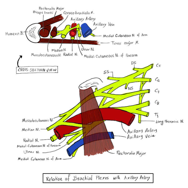
NB: Identification in Cadaver
Find the Axillary Artery (Tube structure that doesn’t stay collapse on pinching unlike veins) at the Axillary space.
- Radial Nerve (Solid cord like structure) can be identified Posteriorly to Axillary Artery running along the shaft of humerus bone.
- Median Nerve can be identified Antero-laterally to Axillary Artery that also run along shaft, and bifurcates if traced backwards to origin (towards neck), which are medial and lateral roots of Median Nerve
- Other Structures hence can be Identified from these by tracing backwards.


thank you!! very helpful post 😁
Thank you for your support