Hip joint is a joint between the head of the femur and acetabulum of the pelvis. The main function of this joint to support the upper body weight in every posture it can be static or dynamic. It also enables the lower limbs to join the the trunk.

Type of joint
- This is a ball and socket type of joint (permits movement in all directions)
- Due to the presence of synovial cavity and fluid it is also known as synovial joint.
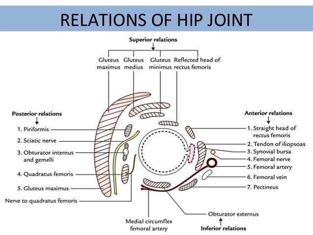
Relations
- Anteriorly- Pectineus muscle, Iliopsoas muscle, Sub tendinous iliac bursa , Femoral nerve and Femoral artery.
- Posteriorly- Superior border of quadratus femoris, Sciatic nerve, Obturator internus and piriformis muscle.
- Laterally- Rectus femoris anterior to iliofemoral ligament , Iliotibial tract and gluteus minimus.
- Superiorly- Gluteus minimus, Gluteus medius and overlying gluteus maximus.
- Inferiorly- Obturator externus inferior to the femoral head.
Ligaments
To further keep the joint in its place ligaments do their work as ligaments are mainly associated in bone to bone joint. So, in the hip joint they are used to strengthen the joint and prevent any kind of displacement during movements. Main three ligaments of the hip joint are:
- Iliofemoral ligament
- Pubofemoral ligament
- Ischiofemoral ligament
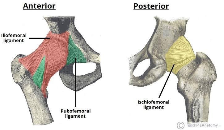
- Iliofemoral ligament- It is triangular shaped ligament which is anterior to the hip joint. It arises from the anterior inferior iliac spine and then bifurcates before inserting into the intertrochanteric line of the femur appears as Y in shape. It is the strongest of the three ligaments.
- Pubofemoral ligament- It is also a triangular shaped joint which is located anteroinferor to the hip joint. It connects iliopubic eminence and obturator membrane to deep surface of iliofemoral ligament by blending with the fibrous membrane which spans between the superior pubic rami and the intertrochanteric line of the femur. It has a triangular shape, and prevents excessive abduction and extension.
- Ischiofemoral ligament- As the name implies it connects ischium and the greater trochanter of femur by attaching deep to the iliofemoral ligament. It has a spiral orientation, and prevents hyperextension and holds the femoral head in the acetabulum.
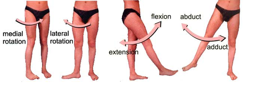
Movements
As we already know that hip joint is a multi axial joint which means it is multiaxial joint and it is capable of performing many kind of movements which include- Adduction, Abduction, Flexion , Extension , External rotation, Medial rotation and Circumflexon.
Their associated muscles are:
- Adduction– Adductor muscles, Pectins and Gracilis.
- Abduction– Gluteus medius and minimus and Tensor facial latae (TFL)
- Flexion– Iliopsoas, Rectus femoris, Sartorius, Pectineus
- Extension– Gluteus maximus, Semimembranosus, Semitendinosus ,Biceps femoris and Adductor Magnus.
- Medial rotation– Same muscles as in abduction but mainly their anterior fibers.
- External rotation– Biceps femoris, Gluteus maximus, Piriformis, Assisted by the obturators, Gemellus and Quadratus femoris.
- Circumflexion– This movement is the addition of all the above movements so it requires all the above muscles and movements work in a equity.
Applied anatomy
- Pelvic fracture- This type of fracture include a bony part fracture of any bone associated with hip joint it can cause pain during certain action and movements and visiting a hospital is very necessary because this condition may cause internal bleeding as a complication in this scenario.
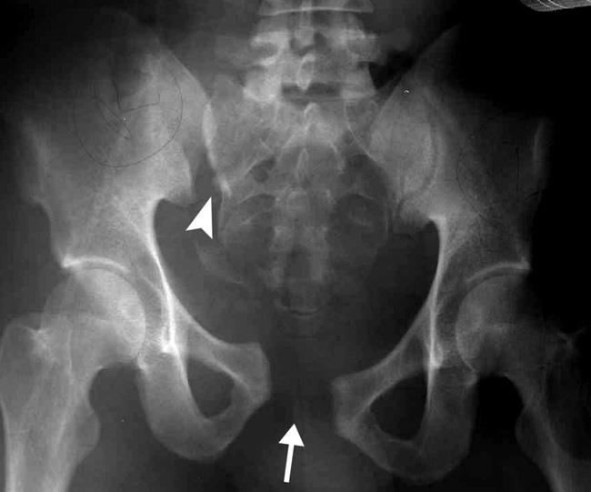
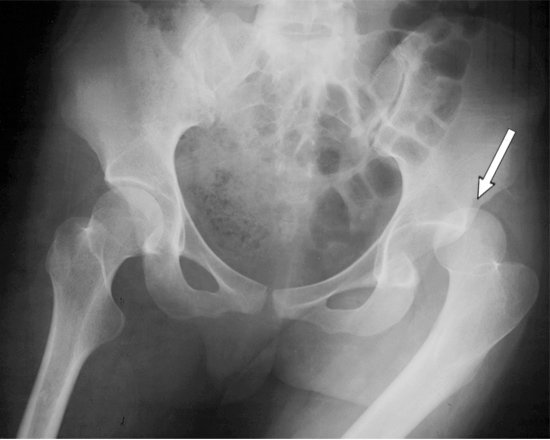
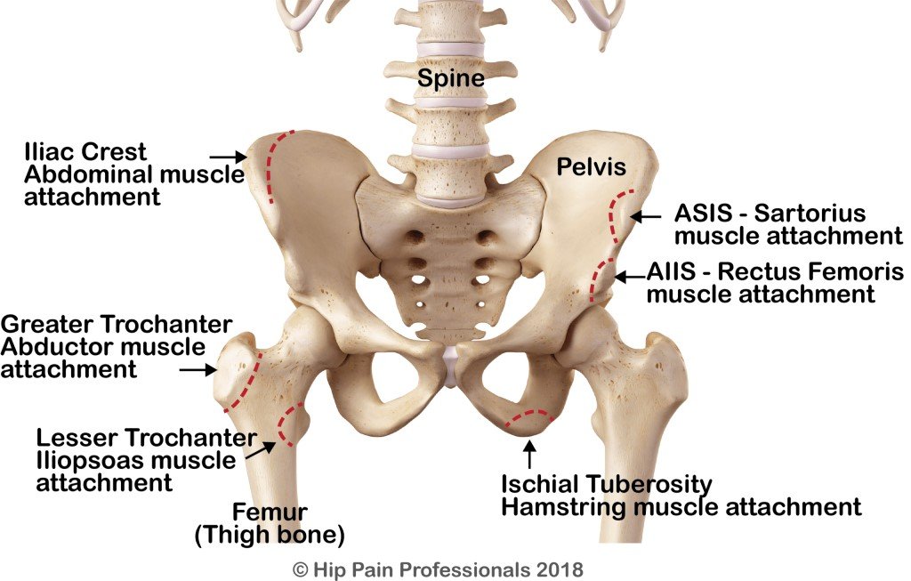
- Avulsion – An avulsion fracture is an injury to the bone in a location where a tendon or ligament attaches to the bone. During some sporty movements such as running, sprinting, kicking a ball, hurdle jumping a small part of tendon or ligament attached to a bone is torn away.
[These are the sites of avulsion fracture where a small piece of bone may dislodge with the muscle that attaches there. Most common sites are depicted on the right and less common sites on the left of the picture]
- Hip joint dislocation- In some injury basically the hip joint is dislocated from its original position and this condition can be treated but this increases the chances of dislocation again in the future. A posteriorly positioned head is the most common dislocation type. Hip dislocations are a medical emergency, requiring prompt placement of the femoral head back into the acetabulum (reduction).
- Posterior dislocations is when the femoral head lies posteriorly after dislocation. It is the most common pattern of dislocation accounting for 90% of hip dislocations.
- Anterior dislocations is when the femoral head lies anteriorly after dislocation. Anterior dislocations are subdivided into two types being inferior (obturator) dislocation and superior (iliac or pubic) dislocation.