Origin – Median Nerve is formed by Lateral cord and Medial cord of brachial plexus
Root value – All the root values corresponding to brachial plexus i.e. C5-C8 and T1

Also known as ‘laborer’s nerve‘ because it gives innervation to all those muscles responsible for complex movements in daily life. It is so called median because it lies in middle of the forearm.
RELATIONS
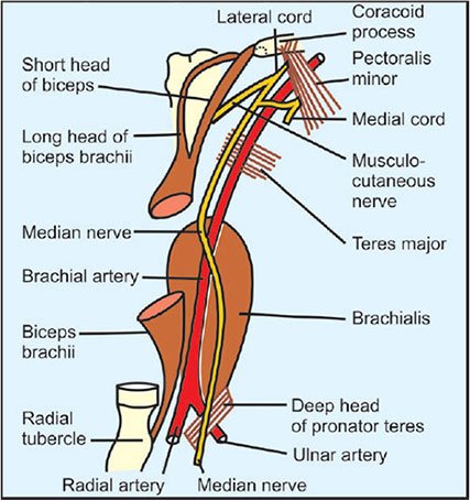
- In upper part of arm median nerve lies lateral to the brachial artery.
- In middle part of arm it crosses the brachial artery from lateral to medial side. By the time it reaches the lower arm, the nerve is medial to brachial artery .
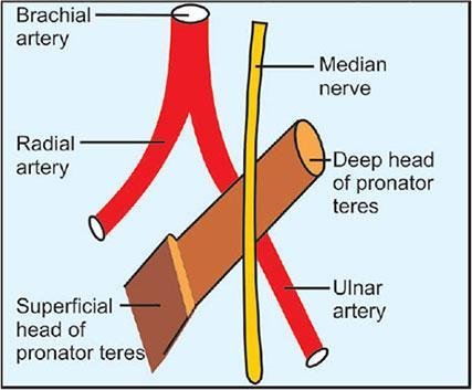
- In cubital fossa it is medial to the brachial artery. But while passing through cubital fossa, median nerve is crossing the ulnar artery and are separated from the ulnar artery by deep head of pronator teres muscle.
COURSE
- Median nerve enters the anterior compartment of arm at the lower border of Teres major.
- In the arm, initially it lies lateral to the brachial artery, then crosses in front of the artery to reach its medial side.
- Enters the cubital fossa where it lies medial to the brachial artery.
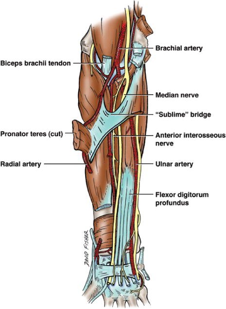
- It leaves the cubital fossa by passing between the two heads of the pronator teres and gives off anterior interosseous nerve.
- In the forearm, it passes behind the tendinous arch of flexor digitorum superficialis and runs downwards deep to the muscle.
- 5 cm. proximal to flexor retinaculum it becomes superficial and lies lateral to the tendons of flexor digitorum superficialis.
- Before entering carpal tunnel it gives off palmar cutaneous branch ( passes superficial to flexor retinaculum) which supplies skin over thenar eminence and lateral part of palm.
- It then enters the palm through the carpal tunnel (deep to flexor retinaculum).
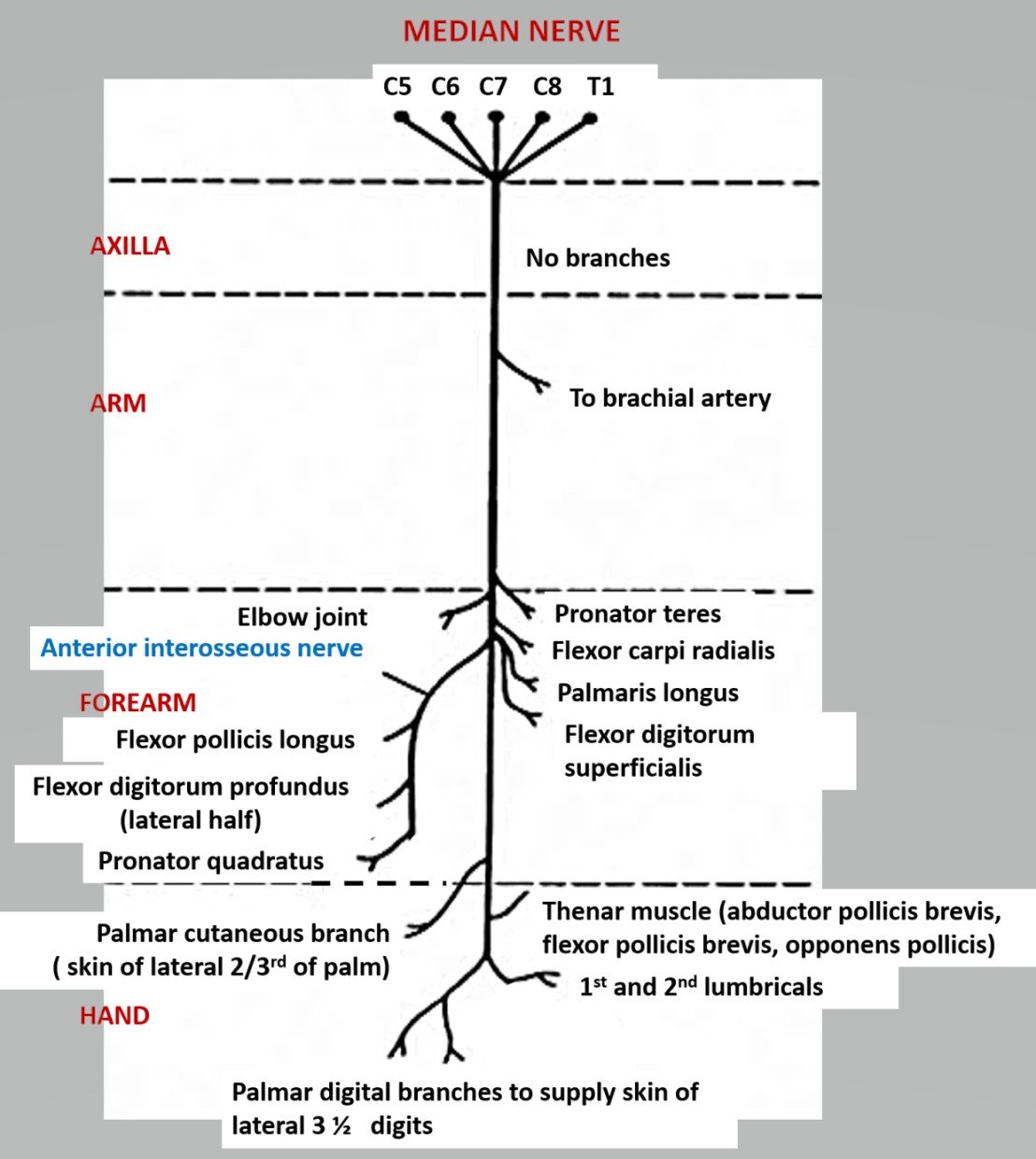
BRANCHES
Arm
Just above the elbow i.e. before entering into cubital fossa it gives branch to pronator teres muscle and a vascular branch to brachial artery.
Cubital fossa
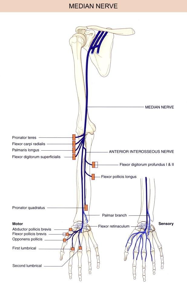
Median nerve here gives muscular branches to supplies all the superficial flexor muscles except Flexor carpi ulnaris
- Flexor carpi radialis
- Palmaris longus
- Flexor digitorum superficialis
Forearm
Anterior interosseous branch supplies 2 ½ muscles –
- Flexor pollicis longus
- Pronator quadrates
- Lateral half of the flexor digitorum profundus.
Palm
Motor branches:
- A recurrent branch that supplies all thenar muscles ( abductor pollicis brevis, flexor pollicis brevis and opponens pollicis) except adductor pollicis.
- Lateral two lumbricals associated with movement of the index and middle fingers
Sensory branches:
- Skin over the palmer surface of lateral three and one-half digits
- lateral 2/3rd of palm of hand
- Distal phalanx of lateral three & half digits on dorsal side.
MOTOR FUNCTIONS
- Thumb flexion and opposition
- Flexion of digits 2 and 3
- Wrist flexion and abduction
- Forearm pronation
CLINICAL ANATOMY
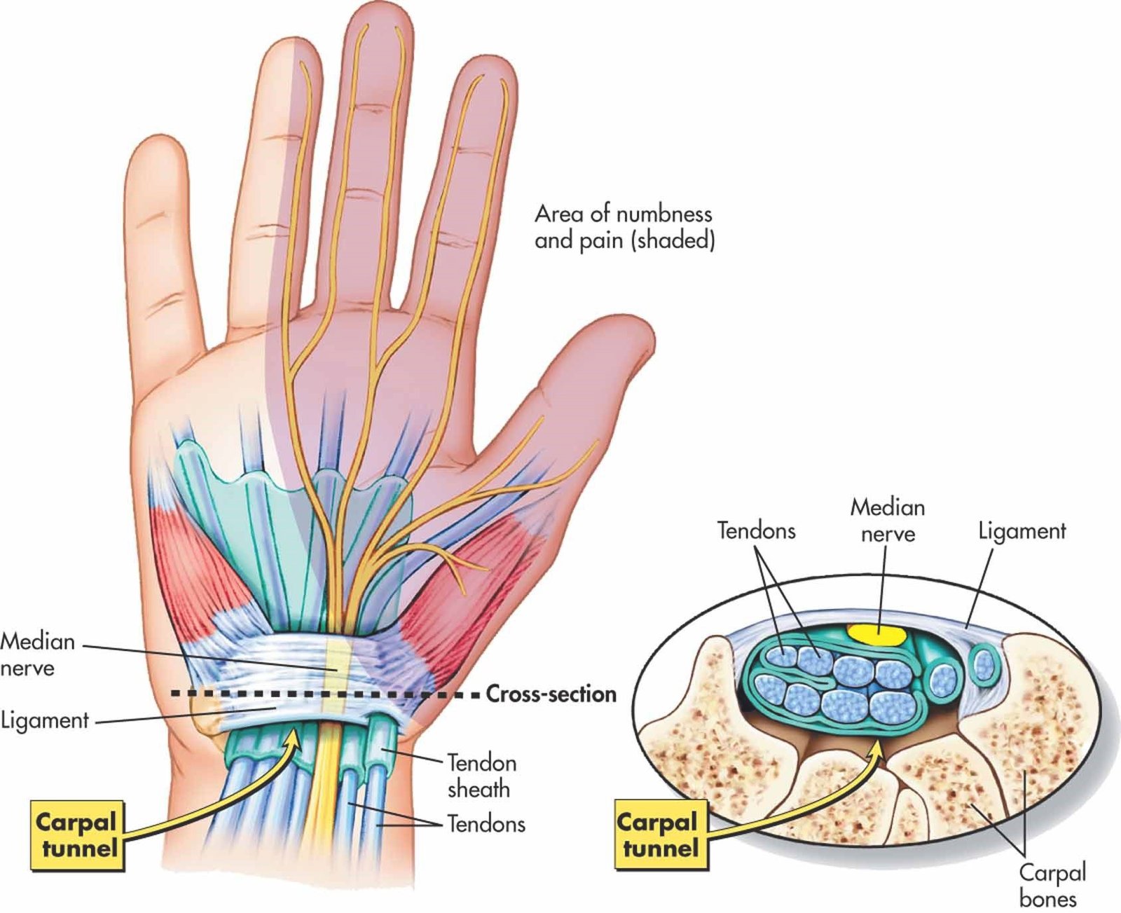
Carpal Tunnel Syndrome –
Due to compression of median nerve in carpal tunnel. It is diagnosed by Phalen maneuver and Tinel sign
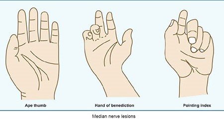
Ape Hand deformity
- Median nerve palsy
- Inability to abduct the thumb. So thumb remains adducted & laterally rotated so that the thumb lies in the same plane as the other fingers. It is due to over action of adductor pollicis (supplied by ulnar nerve).
Hand of Benediction
When patient tries to make fist, then his index and the middle finger remain extended due to paralysis of the flexors of these two digits.
Pointing index/ Oschner’s clasp test
When patient is asked to clasp his hand, index finger fails to flex.


very useful
We are coming with more content. You support will motivate us. Please share this platform with your friends.