INGUINAL CANAL
Inguinal canal is a short passage that extends inferiorly and medially through the inferior part of the abdominal wall. It is superior and parallel to the inguinal ligament. This canal is a pathway for the structures to pass from the abdominal wall to the external genitalia. The inguinal canal begins at the deep inguinal ring and continues for approximately 4 cm, ending at the superficial inguinal ring. In males it conveys the spermatic cords and in females round ligament of uterus.
Any weakness or defect in the abdominal wall leads to herniation.

BOUNDARIES
The inguinal canal is bordered by anterior, posterior, superior (roof) and inferior (floor) walls. It has two openings – the superficial and deep rings.
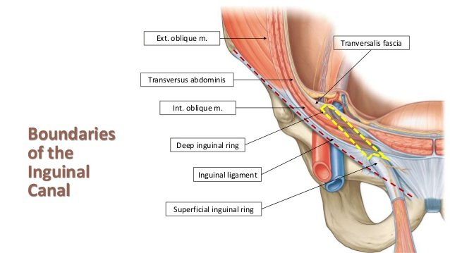
Walls
Anterior wall – Aponeurosis of the external oblique, reinforced by the internal oblique muscle laterally.
Posterior wall – Transversalis fascia.
Roof – Arching fibres of transverse abdominis and internal oblique muscles
Floor – Medial one-half of the inguinal ligament also known as poupart ligament

Rings
Deep inguinal ring – The superficial (internal) inguinal ring is the beginning of the inguinal canal. It has an oval shaped opening in midway between the anterior superior iliac spine and the symphysis pubis above the inguinal ligament. Sometimes referred to as a defect or opening in the transversalis fascia.
Superficial inguinal ring – The superficial (external) inguinal ring is the end of the inguinal canal. It is a triangular-shaped defect in the aponeurosis of the external oblique muscle and lies immediately above and medial to the pubic tubercle.
CONTENTS
The content of inguinal canal are :
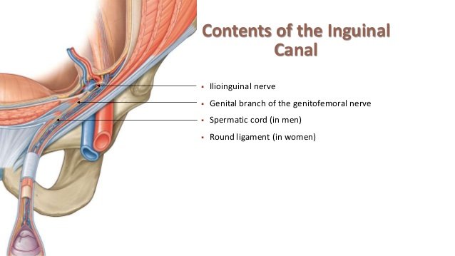
The spermatic cord in men
Ilioinguinal nerve
The round ligament of the uterus
Genital branch of the genitofemoral nerve in women.
INGUINAL TRIANGLE ( Hesselbach ’ s triangle )
Inguinal triangle is present in groin region which is bounded:
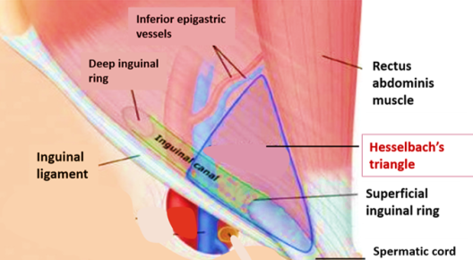
Laterally: Inferior epigastric artery
Medially: Rectus abdominis muscle
Inferiorly: Inguinal ligament.
DEVELOPMENT OF CANAL
During embryogenesis, testicles descend from the posterior abdominal wall and gradually migrate into the scrotal area. This descent or migration of the testicles is guided by a cord-like structure called the gubernaculum. It attaches the inferior pole of the testicles to the developing scrotal sac.
With the descent of the testicles, a peritoneal outpouching called the processus vaginalis follows the testicles to the scrotum. Following the descent of the testicles into the scrotum, the processus vaginalis degenerates. This process of degeneration or obliteration may be delayed, or it may fail completely. Failure of closure of the processus vaginalis leads to development of a number of abnormalities. Peritoneal fluid can travel down a patent processus vaginalis leading to the formation of a hydrocele. Persistent processus vaginalis may increase the risk of development of inguinal hernias.
Source – Wikipedia, Teachmeanatomy
Written by – Dr Divya Ranasaria


HI i am Sadaf and i am in MBBS2 and this topic really helped me out thank you so much!!