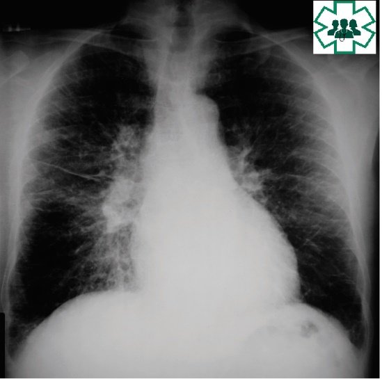
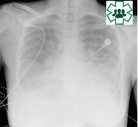
Pulmonary edema is fluid accumulation in the lungs that causes flooding of the alveoli with fluid and the signs include shadow with ill-defined borders, air bronchogram and silhouette sign. This causes severe disturbance of gas exchange across the alveolar surface and can lead to respiratory failure.
There are two distinct radiographic patterns of pulmonary edema: pulmonary alveolar edema and pulmonary interstitial edema.
Causes of pulmonary edema
A. Cardiogenic pulmonary edema: Acute left ventricular failure, Mitral stenosis, Renal failure and Over transfusion of fluids.
B. Non-cardiogenic pulmonary edema: Acute respiratory distress syndrome (ARDS), Renal failure with fluid overload, Iatrogenic fluid overload.
Radiological appearances
Symmetrical, diffuse ‘fuzzy’ shadowing
Especially in the mid and lower zones where the pulmonary venous pressure is highest due to gravity.
Upper lobe blood diversion
Vessels in upper lobe larger than vessels in lower lobe on erect chest
X -ray.
Peribronchial shadowing
Thickened bronchi when viewed end-on. Sign occurring when excess fluid builds up in the small airways causing localised patches of atelectasis. This causes the area around the bronchus to appear more prominent on an X- ray.
Peri-hilar haziness
Hazy shadowing around the hilar regions.
Bat’s wing pattern
In acute pulmonary oedema bilateral or unilateral ill – defined opacities confined to the central (peri-hilar) area of the lungs, extending laterally to stop 2– 3cm before the periphery of the lung called Bat’s wing pattern.

Septal lines
Septal lines are caused by engorgement of the pulmonary interlobular septal lymphatics by fluid, tumour or fibrosis. Found around the periphery of the lungs, extending inwards from the pleural surface.