PALMER SPACES OF HAND
- Pulp space of fingers
- Mid-palmar space and
- Thenar space
To understand these spaces you have to observe the diagrams and understand them carefully.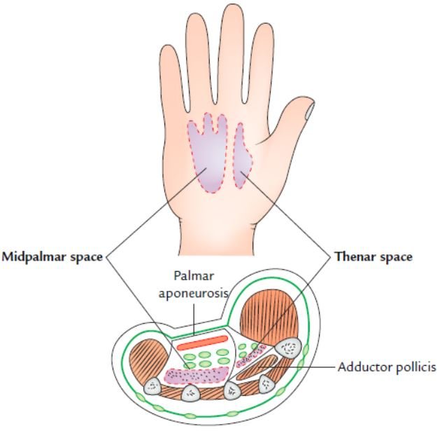
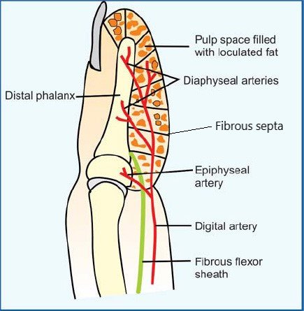
PULP SPACE OF FINGERS
- This space is located behind the skin on tips of finger which continues with distal phalanx.
- There are fibrous septa which communicate to distal phalanx
- This space has got fat and connective tissue which is bound tightly. For this reason, fluid or pus in this area has no space to drain because of tight septa. To treat this, make 2 incisions on either side or pus can infect other spaces too. Infection of this space is known as WHITLOW and is very painful.
- Terminal branches of the digital artery course through the spaces to supply the diaphysis of the distal phalanx.
Boundaries:
- Anteriorly – they are bounded by the skin and superficial fascia
- Posteriorly – they are bounded by the distal 2/3rd of the distal phalanx.
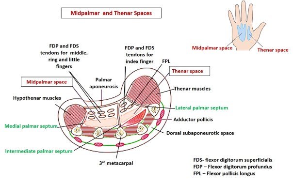
MID PALMAR SPACE
The triangular mid palmar space is located under the medial half of hollow of the palm.
Boundaries:
- Anteriorly – medial part of palmar aponeurosis and flexor tendons of middle three digits (enclosed in ulnar bursa).
- Posteriorly – interossei muscles (3, 4, 5) and 4th and 5th metacarpals.
- Proximally – it communicates with the forearm space of Parona through carpal tunnel.
- Distally – medial three web spaces (subcutaneous spaces in the interdigital cleft) through the medial three lumbrical canals (fascial sheaths of lumbricals).
- Medially – medial palmar septum (extending from the palmar aponeurosis to the 5th metacarpal).
- Laterally – intermediate palmar septum (extending from the palmar aponeurosis to the 3rd metacarpal).
Contents:
- Superficial palmar arch
- Digital nerve and vessels
- Lumbrical muscles
- Long flexor tendon
N.B – Drainage in case of infection: Pus from this space can be drained by incision in the 3rd and 4th web spaces.
THENAR SPACE
As discussed above it is present at lateral aspect.
Boundaries:
- Anteriorly – palmar aponeurosis and flexor tendon of index finger and tendon of flexor pollicis longus (enclosed in radial bursa) and 1st lumbrical.
- Posteriorly – fascia covering adductor pollicis.
- Laterally – lateral palmar septum (extending from the palmar aponeurosis to the 1st metacarpal).
- Medially – it is bounded by the intermediate palmar septum (extending from the palmar aponeurosis to the 3rd metacarpal).
Contents:
- Flexor tendon of index finger and flexor pollicis longus
- Digital nerves and vessels for 1½ fingers.
APPLIED ANATOMY
Infection of mid palmar space: The ulnar bursa is considered as the inlet for infection and lumbrical canals as the outlets of infection in mid palmar space. The pus form this space is drained by incisions in the medial two web spaces.