Femoral sheath
Femoral sheath is a continuation of transversalis fascia of abdomen. It is conical in shape, wider above and narrow below. It surrounds the upper portion of artery, vein and lymphatics but remember it doesn’t enclose the nerve.
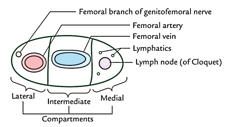
Compartments
As we can appreciate from the diagram, the femoral sheath accounts of three compartments. The order of structures enclosed in the femoral sheath can be remembered by AVL means Artery Vein and lymphatics.
- Lateral – Contains the femoral artery and femoral branch of Genito femoral nerve.
- Intermediate / middle – This compartment contains only a single structure which is a femoral vein.
- Medial – It is called femoral canal, and filled with areolar tissue and contain a lymph node (Gland of Cloquect) which drains glans penis/clitoris.
Functions
Femoral sheath protects the important vasculature of the thigh during the thigh movements and the flexible compartments allow the vascular structure to be separated from each other so they do not compromise each other by pressing on their wall.
Position
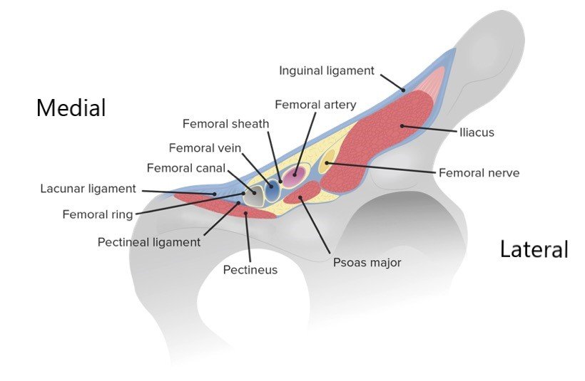
Femoral sheath is located in the femoral triangle which simply tells us that muscles such as the iliacus ,psoas major and pectineus muscles run posterior to the femoral sheath forming the posterior wall of femoral sheath.
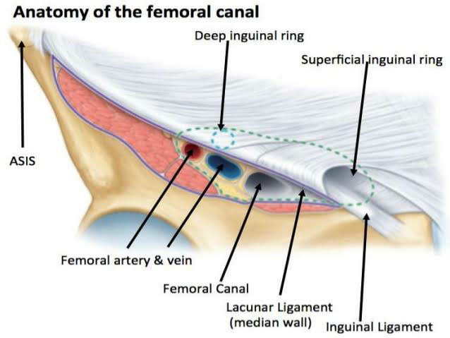
Femoral canal
It is the funnel shaped most medial compartment of the femoral sheath. Femoral canal is an inverted cone shaped canal. It runs from the pelvic cavity to the anteromedial aspect of the thigh and its upper end opens towards abdominal cavity is known as the femoral ring.
Boundaries
- Laterally – Femoral vein
- Medially – Lacunar ligament
- Anteriorly – Inguinal ligament
- Posteriorly – Pectineal line of pubic bone.
Contents
Femoral canal is filled with fat and connective tissue provide cushioning to the main structure present in the femoral canal which are lymphatic vessels which are draining the lymph from deep inguinal lymph nodes, the deep lymph nodes which are also known as the deep inguinal nodes of Cloquet which basically drains the fatty connective tissue, clitoris in female and penis in male.
Clinical significance
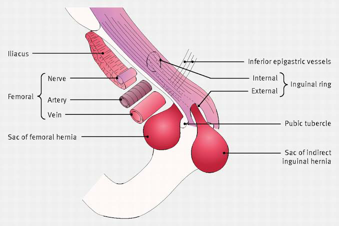
Femoral hernia –
- Femoral hernias are in the list of commonly occurring hernias is the groin region of adults.
- Elderly females are more in danger of femoral hernia because of the wider pelvis.
- Usually the bowel ( small intestine most of the time) protrudes through the femoral ring into the femoral canal.
- Two conditions can be form in this type of hernia. If the intestines are stuck but still has the blood flow, is called as incarcerated femoral hernia. And if the intestines are stuck with the loss of blood blow due to compression is known as strangulated hernia.
- Femoral hernia has following coverings:
- Femoral septum
- Anterior wall of femoral sheath
- Cribriform fascia
- Surgical reduction of femoral hernia: Lacunar ligament has to be divided to relieve strangulation.
#Know more about lower limbs from “THIS LINK“.
#We Partnered with Achievable.me. Check out their website for awesome study content.


Very clear anatomical details and applied anatomy of femoral sheath and femoral canal with diagrams.
Thanks a lot doc!! All credits go to our talented author Dr. Mohit Naruka..
Thank you team☺
Thank you so much Doc